abnormal thyroid cancer ultrasound colors
Ultrasound uses soundwaves to create a picture of the structure of the thyroid. An abnormal growth of thyroid cells that forms a lump within the thyroid.

Pin On Shitovidka
A thyroid ultrasound is a safe painless procedure that uses sound waves to examine the thyroid gland.

. The normal thyroid gland is located in the anterior lower neck between the thyroid cartilage and the thoracic inlet. It is generally normal unless there is too much color which would have been mentioned in the report. It can be used to help diagnose a wide range of medical conditions affecting the thyroid gland including benign thyroid nodules and possible thyroid cancers.
Color and Power Doppler ultrasound failed to show significant vascularity within the affected area lesion in the right lobe. For example nodules that do not have smooth borders or have little bright white spots micro. This nodule shown in red comprises about 80 of the thyroid tissue shown in yellow in this particular area of the thyroid.
Ultrasound color and grayscale images pictures Ultrasound Color images. Use MFI color imaging if possible. It measures 057 x 061 x 069cm.
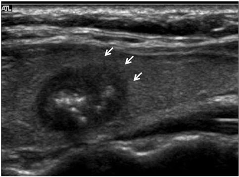
Ultrasound showed a well-defined rounded isoechoic solid nodule in the upper pole of right thyroid lobe. Color on your thyroid ultrasound means that color doppler was applied and blood flow was detected. Recommend FNA for sampling if 1 cm in greatest dimension and high suspicion sonographic pattern estimates a.
What is the red and blue on a thyroid ultrasound. To determine whether color Doppler interrogation of a thyroid nodule can aid in the prediction of malignancy. Strongly recommend ultrasonic examination of thyroid and cervical lymph nodes if thyroid nodules.
It is same as normal sound in its physical properties but humans cannot hear it. A solid one is more likely to have cancerous cells but youll still need more tests to find out. Pin On Sono Ultrasound Pin By Dr Abuaiad On Lymphatics Diagnostic Medical Sonography Nuclear Medicine Sonography Pin On Medical Imaging Pin On Uh Pin On Thyroide Pin On Shitovidka Pin On Tirads Us Thyroid Pin On Superficial.
You would only see normal thyroid tissue. Other links to color or gray scale images will be included here later. These images are examples of pathology I detect with sonograms.
Ultrasound is a sound wave with a frequency higher than 20kHz used to look at organs and structures inside the body. A thyroid function test was done and confirmed T4 is elevated. Doctors typically evaluate thyroid nodules using ultrasound scans.
To determine whether color Doppler interrogation of a thyroid nodule can aid in the prediction of malignancy. Ultrasound devices range with frequencies from 20kHz to several gigahertz. An ultrasound may show your doctor if a lump is filled with fluid or if its solid.
Thyroid is a gland that serves several functions that affect the well being of the body. For papillary thyroid cancer the 20-year survival after surgery is around 99. Certain characteristics of thyroid nodules seen on an ultrasound are more worrisome than others.
SerhiiBobyk iStockGetty Images Plus Getty Images. Ultrasound imaging of the thyroid gland shows markedly hypoechoic lesions in the right lobe. Color Doppler showed increased vascularity while the rest of the thyroid showed normal vascularity.
While most thyroid nodules are non-cancerous Benign 5 are cancerous. She was then referred to me for ultrasound. We obtained color Doppler images of thyroid nodules undergoing sonographically guided fine-needle aspiration.
Use Color flow imaging of abnormal appearing lymph nodes. VIEWPOINT drawing to illustrate any abnormal lymph nodes should be created for the following patients- Pre-surgical Thyroidectomy patients or known thyroid cancer patients. We obtained color Doppler images of thyroid nodules undergoing sonographically guided fine-needle aspiration.
If you looked at other parts of the thyroid however you would not see the nodulem. The 3 mm nodule is likely of. Undefined 41 years experience.
A common imaging test used to evaluate the structure of the thyroid gland. Hypervascularity is a typical finding in people with underlying autoimmune thyroiditis hashimotos or graves disease. After thyroidectomy the local inflammatory response results in proliferation of fibrofatty connective tissue which fills the dead space made by surgery There is also displacement of the strap muscles the carotid sheath structures and the cervical esophagus.
Abnormal thyroid cancer ultrasound colors Friday February 18 2022 Edit. There are certain factors that make a nodule suspicious for thyroid cancer. The most prevalent form of thyroid cancer is papillary thyroid cancer 75-80 followed by follicular 10-20 medullary 3-5 and anaplastic 1-2 thyroid cancers 2 26.
The color Doppler appearance of each nodule was graded from 0 for no visible flow through 4 for extensive internal flow. The survival rate for thyroid cancer in general is better than for other forms of cancer. Thyroid hypoechogenicity at ultrasound is a characteristic of autoimmune thyroid diseases with an overlap of this echographic pattern in patients affected by Graves disease or Hashimotos thyroiditis.
The color Doppler appearance of each nodule was graded from 0 for no visible flow through 4 for extensive internal flow. Hypoechoic thyroid nodules appear dark relative to the surrounding tissue. Aim of the present paper was to study the thyroid blood flow TBF by color-flow doppler CFD an.
The ultrasound will also show the size and number of nodules on your thyroid. Seroma hematoma abscess tumors. What Does Colour on an Ultrasound Means.
2015 American Thyroid Association ATA Management Guidelines. They use this information for surgical planning. If there was an abnormal finding in the thyroid ultrasound the patient was referred to an endocrinologist and after clinical and laboratory evaluation fine-needle aspiration FNA biopsy was done if required.
Patients and methods. Can you detect thyroid cancer in ultrasound. Red and blue denote.
48k views Answered 2 years ago. Buy 2021 Quality Abnormal Thyroid Cancer Ultrasound Color Doppler directly with low price and high quality. The first links in each row here correspond to ultrasound color post-processed images.
The thyroid gland was evaluated for any nodules following carotid Doppler ultrasound in 290 patients. Ad Learn more about the signs that may reveal you have an Issue that need attention. Thyroid nodules are common medical and surgical problems They can be evaluated by many techniques including physical examination fine needle aspiration and imaging Even though thyroid malignancy may be detected in 5 of thyroid nodules early diagnosis and treatment are recommended because of nodules slow progress and long.
The hypoechoic thyroid lesion shows irregular borders and is seen to infiltrate along the long axis of the affected lobe. For Adult Patients with Thyroid Nodules and Differentiated Thyroid Cancer. What are white spots on thyroid ultrasound.

Pin On Thyroide

Pin On Thyroide

Pin On Sono Ultrasound

Pin On Thyroide

A Gallery Of High Resolution Ultrasound Color Doppler 3d Images Thyroid Thyroid Ultrasound Thyroid Ultrasound

Pin On Sonography

Pin On Superficial

Pin On Sono

Pin On Superficial

Pin On Tirads Us Thyroid

Pin Na Doske Medical Stuff

Pin By Dr Abuaiad On Lymphatics Diagnostic Medical Sonography Nuclear Medicine Sonography

Pin On Thyroide

Pin By Dr Abuaiad On Superficial Ultrasound Technician Ultrasound Sonography

Pin On Superficial
Pin On Medical Imaging

Pin On Dr
Pin On Abd 300 Mod 4 Ultrasound

Pin On Thyroide